Transcript
the Oregon State University honey lab
presents dissecting for trach
mites to determine if a bee is infested
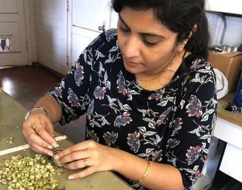
with trach mites one must dissect the
bee to view tracheal
tubes place the bee on its back with
forceps remove the head and first pair
of legs
pull on the collar of the thorax and
remove the collar may be brittle and
break apart so the technique may take
practice trachea should be attached to
the collar piece or they may still be
attached to the B
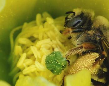
body this is an adult trul Mite and an
egg that have been removed with forceps
from the the trachea they are small
white and football-shaped
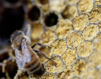
this is a tracho tube with no trach
mites this is a mild infestation of
trach
mites this is a moderate
infestation this is a heavy infestation
of tracheo
mites severe infections may cause dark
scarring of the
trachea however scarring can occur with
different levels of
infestation
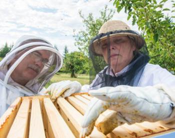
Summary
Knowing the levels of tracheal mites in your apiary and understanding the effects of their presence will enable you to make an educated decision on your treatment plan. In this video, we demonstrate the dissection of a honey bee worker for tracheal mite infestation.
Catalog - EM 9145